News & Updates
News & Updates
Development of trimer atom based gas field ion source for ion microscopy
- Writerkrissadmin
- Date2018-07-11 00:00
- Hits18361
Development of trimer atom based gas field ion source for ion microscopy
- Advances in ion beam technology with reverse use of oxide film -
# In the world of microscopy, quality depends on beams such as light, electrons, and ions. How such sources are utilized determine the performance of microscopes. Higher-resolution images can be obtained when beams are concentrated to a small area and then emitted.
The Korea Research Institute of Standards and Science (KRISS, President Sang-Ryoul Park) developed novel ion beam* technology for helium ion microscopes, which are recognized as next-generation microscopy.
* Ion beam: A charged particle beam comprised of ion. An ion microscope uses an ion beam as its source.
The research team led by In-Yong Park of the Photon and Electron Instruments Team at KRISS designed an ion source device*, and succeeded in generating ion beams with a trimer atom probe.
* Ion source device
: A device that generates ion beams by ionizing inactive gases surrounding the probe when a strong positive voltage is applied after introducing inactive gases such as helium, neon, and xenon
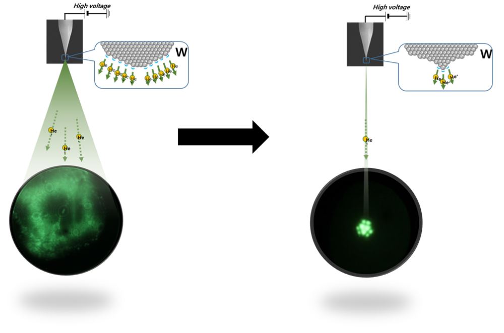
▲ Comparison of multi-atom ion source (left) and trimer atom ion source device (right)
- The multi-atom probe before etching (left) emits ion beams in various directions over a large area,
while the proposed probe (right) emits high-brightness ion beams over a very small area (three atoms).
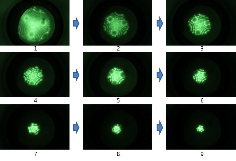
▲ Fabrication of the trimer atom probe using the ion source device
- Etching of the probe to the size of three atoms using oxygen in the insulating layer
Helium ion microscopy not only offers sub-nanometer resolution, but is also suited for precision manufacturing of dimensions less than 10 nanometers. For this reason, helium ion microscopes have diverse applications in various fields including nanoprocess technology, materials science, and biology.
Ion sources must be precisely designed to achieve high resolution in helium ion microscopy. The area over which ion beams are emitted is made smaller by sharpening the probe tip. The key to a sharp probe tip is to leave a minimal number of atoms at the tip.
High-performance ion beam technology is very important in ion microscopy, but commercial microscopes with trimer atom probes are made by only a handful of international companies due to their technical complexity.
A major element that interferes with probe tip fabrication is the oxide film. When the probe is exposed to air before entering the vacuum environment, an oxide film forms on the tungsten surface. In the past, this oxide film was removed through heat treatment of the probe.
Park and his team succeeded in developing a trimer atom probe by making use of the oxide film, which was previously removed by applying heat to the insulating layer. The team used the oxygen in the insulating layer for etching, allowing the probe to have a sharper tip.
The significance of this study is that it lays a foundation to develop more advanced microscopes without relying on exports. Since heat treatment is no longer necessary, ion beams can be generated more conveniently and efficiently.
Park said, “The ion beam technology reduces the number of steps involved compared to existing equipment. It is expected to significantly enhance the competitiveness of the local market, which is dominated by imports. Our ultimate goal is to develop a single atom probe that uses only a single atom to achieve three times greater brightness.”
Supported by the Nanotechnology Development Project of the National Research Foundation of Korea, the study was published online in the June issue of the leading journal, Ultramicroscopy.
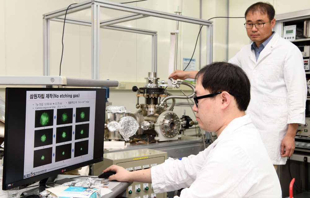
▲ Park (back) and his team performing the trimer atom etching test
QUICK MENU
QUICK MENU 원하시는 서비스를 클릭하세요!
등록된 퀵메뉴가 없습니다.