News & Updates
News & Updates
Perivascular health an important factor in atherosclerosis
- Writerkrissadmin
- Date2019-08-21 00:00
- Hits12355
Perivascular health an important factor in atherosclerosis
- First demonstration of perivascular deterioration in atherosclerosis-
- Potential for atherosclerosis prediction and treatment based on perivascular changes -
# Although atherosclerosis is a major health threat, we tend to think of it as a disease that causes blockage within blood vessels. Atherosclerosis refers to the phenomenon in which cholesterol or triglycerides build on the innermost wall of blood vessels, causing blood vessels to become narrower and lose elasticity. However, a recent study has revealed that atherosclerosis also affects the external wall of blood vessels. This means that atherosclerosis can no longer be seen as a disease that only occurs within blood vessels.
Researchers at the Korea Research Institute of Standards and Science have discovered that atherosclerosis not only causes deterioration within the blood vessels but also damages external walls.
The team led by Dr. Se-Hwa Kim, principal research scientist at the KRISS Center for Nano-Bio Measurement, successfully uncovered the mechanism of perivascular adipose tissue* browning, irregular agglomeration and fibrosis according to the progression of atherosclerosis. The findings of this study could lead to the development of a new diagnostic technology to predict vascular health within blood vessels based on changes in the external wall.
? Perivascular adipose tissue (PVAT)
The outermost tissue layer surrounding blood vessels. Until now, PVAT was only seen as a support structure in blood vessels and was frequently ignored. Recently, with the discovery of its important role in maintaining vascular health, active research is being conducted on this overlooked area.
Atherosclerosis is a major cardiovascular disease that has no symptoms in the early stage, its effects only becoming noticeable when the blood vessels have narrowed to 50% of their original width. As atherosclerosis can lead to fatal diseases such as myocardial infarction and stroke, early detection is of utmost importance.
Atherosclerosis begins with changes in the internal walls of blood vessels. Thus, most studies have focused on what happens inside the vessels, with limited interest in the external wall. The external walls of blood vessels, composed of adipocytes, immunocytes and fibroblasts, are not easy to observe anatomically, and were only regarded as mechanical support for the actual blood vessel.
Recently, there has been a surge in interest in these blood vessels with the introduction of the hypothesis that perivascular adipose tissue plays a crucial part in maintaining vascular health. However, this was mainly attributed to an effect on the endocrine system via hormones secreted from the perivascular adipose tissue, and no direct link between the external walls of blood vessels and atherosclerosis was proposed. Perivascular adipose tissue is challenging to study anatomically as it is soft, without fixed form, and cannot be stained as it is made mostly of fat cells. In fact, it was often cut away and discarded in studies of blood vessels.
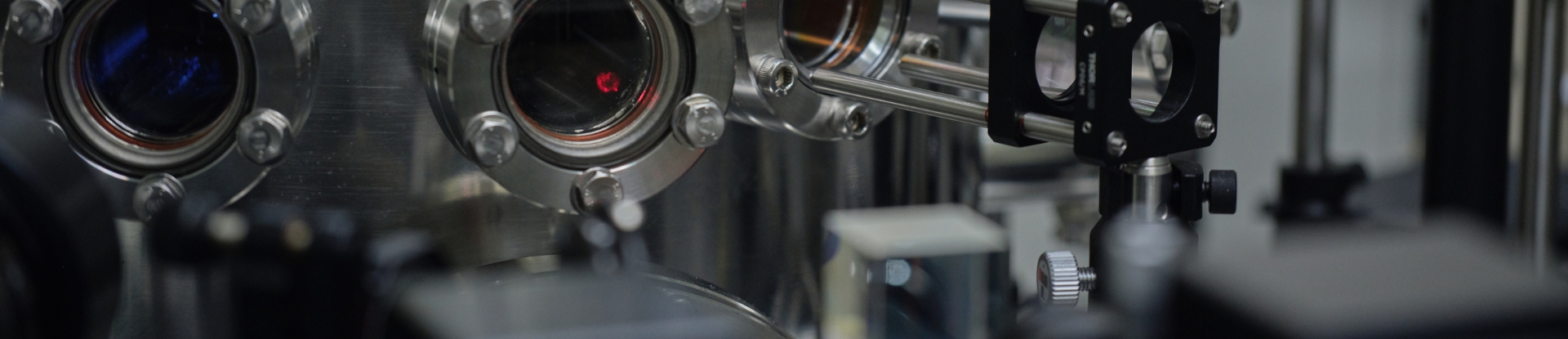
Dr. Se-Hwa Kim’s team at KRISS developed a core technology for obtaining 3D images of perivascular adipose tissue without conventional staining by using a multimodal nonlinear optical microscope. The team successfully performed close up studies of the fat, collagen and elastin making up the blood vessel, keeping the vessels intact without the use of chemical processing.
Through these observations, the team discovered that the external walls of blood vessels also change according to the progression of atherosclerosis. In the early stage of the disease, the level of brown fat in the perivascular adipose tissue increased to raise energy consumption leading to protection of the blood vessels from atherosclerosis. However, with time, the perivascular adipose tissue closer to the atherosclerotic plaque within the blood vessels deteriorated, losing its protective function. The team found that fibrosis of the perivascular adipose tissue was induced by transforming growth factor-beta (TGF-β)*, breaking down the regular array of adipocytes and causing PVAT deterioration.
? Transforming growth factor-beta (TGF-β)
A signaling protein that plays many roles in the body. It is known to affect embryonic formation, differentiation of mature cells and even cancer cell activity.
“It can be interpreted as the external wall of blood cells going beyond the simple mechanical support, to perform a crucial function in controlling diseases within the blood vessel,” says KRISS principal research scientist Dr. Se-Hwa Kim, adding, “there is potential for its use in assessing vascular health within the blood vessel through changes in the exterior. Going beyond that, it may even be used to develop new drugs and therapy targeted at perivascular adipose tissue related disease mechanisms.”
This study was published online in the Proceedings of the National Academy of Sciences (IF 9.661) on August 19th (local time).
○ Images illustrating the study
![그림입니다. 원본 그림의 이름: 크기변환_[첨부1-1] 동맥경화의 진행에 따른 혈관주변지방조직(PVAT)의 변화.jpg 원본 그림의 크기: 가로 1675pixel, 세로 939pixel](/ease_src/crosseditor/binary/images/000002/20210924162128707_R8RV9DS8.jpg)
▲ Changes in the external wall with the progression of atherosclerosis
- (A) Normal perivascular adipose tissue (PVAT).
- (B) Browning of PVAT in the early stage to protect the blood vessel from atherosclerosis.
- (C) PVAT deterioration and loss of function in atherosclerotic plaque regions within the blood vessel.
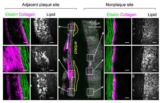
▲ Result of atherosclerotic blood vessel imaging
- External wall of blood vessel adjacent to atherosclerotic plaque (left) has increased collagen showing fibrosis, and abnormal fat tissue forms.
- On the other hand, the external walls of blood vessels without atherosclerotic plaque (right) appear normal.
![그림입니다. 원본 그림의 이름: [첨부2] 비선형광학현미경으로 이미징한 혈관의 모습-합본.jpg 원본 그림의 크기: 가로 700pixel, 세로 700pixel](/ease_src/crosseditor/binary/images/000002/20210924162128731_WDT5VM1T.jpg)
▲ Blood vessels imaged using non-linear optical microscope:
Various tissues composing the blood vessels can be distinguished without chemical staining.
- Fat (Bright yellow or red), collagen (purple), elastin (green)
○ What are its applications?
1. Medical diagnostic technology– precision in atherosclerosis diagnosis
Early and precise diagnosis of atherosclerosis, which is caused by inflammatory response in the blood vessels, has been difficult with conventional medical imaging technology. The present technology moves away from existing atherosclerosis diagnostics, which focus on changes within the blood vessel, to infer vascular health from changes in the external walls of blood vessels.
Going further, through precise measurement and disease prediction research in clinical and pre-clinical studies, combined with imaging technology focusing on changes in perivascular tissue, this technology can be developed into one for assessing inflammatory response levels within blood vessels, as well as blood vessel stability.
2. New drug development– From preventive measures to treatments
The external wall of blood vessels plays an important role from the onset of atherosclerosis to its progression. Most notable is the fact that PVAT browning accelerated in the early stage of atherosclerosis. This could be the key to new preventive technology for vascular disease. PVAT browning continues for some time, until fibrosis occurs around the atherosclerotic plaque, causing the loss of its protective function. Thus, a major regulator for PVAT fibrosis could become a new therapeutic target.
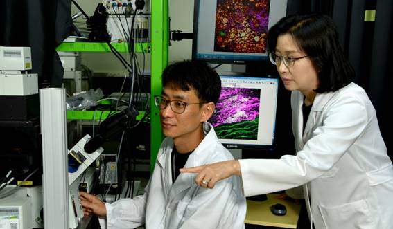
▲ Dr. Se-Hwa Kim (right), principal research scientist at KRISS Center for Nano-Bio Measurement is observing blood vessels using a non-linear optical microscope.
QUICK MENU
QUICK MENU 원하시는 서비스를 클릭하세요!
등록된 퀵메뉴가 없습니다.